VIDEO


ARTICLES
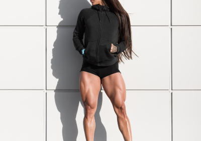
One of the most important running muscles is the quadriceps - the large muscle at the front of the thigh. It has been scientifically proven that regular strength training improves workout performance and reduces the risk of injury, as well as strengthens your back, and leg muscles, making your running more productive.
The quads, also known as the quadriceps muscle, is a group of muscles located at the front of the leg above the knee. It is one of the largest and strongest muscles in your body that consists of four heads: fastus medialis (medial wide), vastus intermedius (intermediate wide), vastus lateralis (lateral), and rectus femoris (straight thigh muscle). Almost every action performed by the legs involves two or more heads of the quadriceps. The quadriceps muscle plays an important role at the beginning of the movement (quick start) and during ascent and descent in elevated positions - which is why you may experience painful sensations in this area of the leg after an intense uphill workout.
According to researchers at the University of Delaware, strong quadriceps act as cushioning and can protect the knees and the entire musculature of the hip from common running injuries. Often many runners are injured because they have weak and underdeveloped quadriceps, so it is especially important to strengthen them.
By training and strengthening the quads, you can get the following benefits when running:
Increased speed. Strong quadriceps help you tackle steep hills and climbs more easily and make pushing off the ground more powerful, resulting in increased speed.
Reduced injuries. According to research, poorly developed quadriceps are often associated with many running injuries. For example, a common injury such as the 'runner`s knee' results from weakness in these muscles, which are unable to stabilize the knee joint during the movement.
Increased endurance. Strengthening the quadriceps muscle will make your running more effortless and efficient, which contributes to increased running volume and endurance.
Training
The quadriceps occupy 70% of the muscle mass of the leg, so it is their development that is fundamental in leg training. The main exercise for developing the quadriceps is the squat. But, for beginners, at the first stages of training, it is better to start with leg curls sitting in an exercise machine, leg press, and hyperextensions to strengthen the lower back, to avoid injuries, in order to prepare the base for the heavy squats. It is a good idea to include squats in your training plan after about six months of training.
Read more

Hamstring muscle training is often neglected, but for most athletes, however, it is very important because strong hamstrings help avoid many injuries.
They are a group of powerful muscles that extend from the pelvis to the knee at the back of the upper leg. Their two main roles are to extend the leg (pull it back) and bend the knee, movements we use both in mountain walking or running and in everyday life. We engage and overuse them more than we realize, which creates tension and can lead to acute or chronic injuries due to overuse. For example, when you run or hike up a mountain, it may seem like you are primarily using your quadriceps, but you are also straining your hams, especially if you are overzealous or taking a big step to navigating over rocks or roots.
These muscles consist of three parts: the semi-tendon muscle, the biceps femoris muscle, and the semitendinosus muscle. Together they form a large group of muscles that work at the base of the thigh. Their main function is to bend the leg at the knee joint.
Before performing any strength exercises, it is important to warm up and stretch the hamstring muscles well, because the muscles are often in a contracted condition.
Training
It is better, to begin with, a 5-10 minutes light workout. This can be cycling, running, etc. Such workouts are good for warming up before stretching, after which you can proceed to the main workout. Training cool muscles is not a good idea.
Wise advice for beginner athletes who want to train with extra weight - use a lighter load and more reps at first to gradually develop muscle strength.
There are many exercises that do not directly target your hamstrings, but where they help your hamstrings, such as pull-ups, lunges, and squats. However, to develop them well, it is also important to do some isolation exercises where you focus directly on your hams. To help you on your way, we decided to list the most important exercises below.
Hamstrings Compression helps relieve pain and tightness.
Stretching relieves stress and helps strengthen the ligaments in the back of the knee as well as the hamstrings.
Quadriceps stretching with support. This exercise will strengthen your hams instead of your quadriceps. It also improves knee mobility.
Leg lift with resting on arms. Lifting one leg is aimed at pumping the glutes, as well as strengthening the hamstrings.
Wall Squats. Such squats will strengthen your knees and also reduce pain if any. The exercise works all the muscles that are connected to the knees.
Lunges also engage your hamstrings, glutes, inner thigh muscles, and calves. The strength of these muscles determines the strength of your knees. The stronger they are, the more tension they will absorb, thus preventing injury and pain.
Exercises with a roller are a perfect way to massage and relax your muscles.
Read more
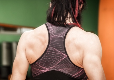
The trapezius muscle is a crucial straight broad muscle that is located in a superficial position, in the back part of the neck and the upper back. The trapezius muscle has the shape of a triangle, the base facing the spinal column, and the top - to the acromion of the scapula. These kinds of muscles on both sides of the back together are trapezoidal.
There are three parts of it:
Upper part: Once the spine is fixed and raises the scapula. With a fixed shoulder blade, it straightens the head and neck.
Middle part: When the spine is fixed and the head brings the scapula to the spine. With the contraction of all parts of the muscles, the adduction of the shoulder blades also occurs.
Low part: It downs the shoulder girdle when the spine and head are fixed.
The upper trapezius muscles are trained by raising and lowering the shoulders with weights in the hands (performing shrugs). The lower part is trained by bringing the shoulder blades of the back under load.
Training.
Band Seated Row. Sitting on the floor with your legs extended loop an elastic band around your soles and hold one end in your right hand. Squeezing your shoulder blades and making a twist to the right with your torso, pull the band toward your waist. Then repeat the action to the other side.
Planche. It is an advanced gymnastic exercise. You should have a high level of fitness preparation and strong hands. It is the skill in which your body should be parallel to the ground supported by hands and arms with your legs raised.
S-Leg Pushup. Put your hands and toes on the mat. Keep your arms straight, but don't lock your elbows. Align your feet making the straight line with your arms and core. Inhaling, bend your elbows and lower your chest to the mat slowly. Straighten your arms to lift your core up, exhaling, at the same time lifting one leg off the ground.
P-up Variation. Put your hands and toes on the mat. Keep your arms straight, but don't lock your elbows. Align your feet making the straight line with your arms and core. Inhaling, bend your elbows and lower your chest to the bench slowly. Straighten your arms to lift your core up, exhaling.
Read more
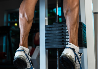
Building up the calf muscles and make your legs beautiful is quite difficult, but possible. It is difficult because the legs are used to constant strain (usually people walk a lot). And it becomes possible thanks to the proven exercises, which can effectively train this group of muscles. In this article, we will take a detailed look at the anatomy of calves, their functions, and exercises that you can do to help make them stronger.
The calf muscle is the most superficially located muscle of the lower leg that crosses two joints: the knee and ankle. Like the biceps of the shoulder, it consists of two heads: medial (located closer to the inside of the tibia) and lateral (located closer to the outside of the tibia). It is interesting to note, that about 5.5% of Japanese and about 3% of people of other nationalities may have the 3rd head of the calf muscle, which attaches from above between the fixation sites of the medial and lateral heads, and may connect from below either to the lateral head (less often) or to the medial head (more often).
The primary function of the calf muscle is to lift the supporting leg, which causes a person to move forward along with flexion of the knee and ankle joints. Running causes a lot of stress on the calf muscles that is why many sprinters face the problem of calf pain.
There are also other functions of the calf muscle.
The calf muscle is involved in the plantar flexion of the foot, which occurs during walking, running, or cycling;
It is also activated during flexion of the leg at the knee joint;
It participates in the supination of the foot (its outward rotation);
The calf muscle takes part in the stabilization of the knee and ankle joints and also helps to keep the balance in the upright position of the body.
There are many reasons that can cause calf pain. And to prevent this unpleasant situation, the following rules should be followed:
running at a comfortable pace;
warming up before running and stretching after training;
comfortable shoes suitable for running;
a smooth transition from running to stopping after a step;
drinking plenty of water after the workout;
a warm shower/bath and a massage to relax the muscles.
The reasons why the calf muscles are not as developed as you would like them to be could be the following:
too much training aimed at working out this muscle group;
not enough exercise;
the wrong set of exercises.
It is necessary to choose the right exercises for the calf muscles. It must be noted that the surface muscle is worked out in a standing position, and the cambalic muscle in a sitting position. To train as effectively as possible, it is necessary to load both muscles.
And the last, but not the least advice: do not try to achieve serious results by performing endless repetitions, such as deadlifts. The main secret of how to pump up the calf muscles is heavy training with weights. It is recommended to train your calves no more than 2 times a week.
Read more

In bodybuilding and fitness, the deltoid muscles play a special part. Despite the fact that the muscle area belongs to small groups, it is second only to the biceps in terms of training frequency. The reason lies not only in the importance of the deltoid muscles in the anatomy but also in improving the aesthetics of the figure. Understanding the functions and features of the deltoid bands allows you to maximize the effectiveness of the training process and significantly reduce the injury risk.
The deltoid muscles consist of three heads (bundles):
The anterior head (anterior bundle) attaches to the humerus and is responsible for lifting the arm forward.
The middle head (lateral bundle) is attached to the acromion of the scapula and allows the arm to be lifted sideways.
The posterior head (posterior bundle) is attached to the scapula and allows the arm to be moved backward.
The deltoid muscle covers the shoulder joint. The muscle is thick, triangular in shape, with the base up and the apex down. It consists of large muscle bundles, which fan-like converge at the apex. It starts from the clavicle and scapula and attaches to the deltoid tuberosity of the humerus.
In training the deltoid muscles, it is very important to pump all three bundles equally. This will protect the shoulder joint from injuries.
The deltoid muscle bundles have different functions, so you cannot work them all with just one exercise: you must include at least three movements in your workout.
You must correctly select exercises for pumping the front, middle and rear bundles. Choose one exercise from each category and add them to your workouts.
Shoulders should be trained no more than twice a week (once is enough for beginners), this will allow the muscles to fully recover for the next workout.
Here is some basic exercises for deltoids:
The army bench press or barbell/dumbbell press in standing or sitting position (middle, front).
Bench press from behind the head while seated (middle, front).
Chin-up or 'broach' barbell pull (middle, front).
Lee Heini pulls (back, middle).
Arnold press (front, middle).
Read more